What it is: A safe and painless medical imaging technique that uses high-frequency sound waves (not radiation) to create real-time, live images of internal organs and soft tissues. It’s commonly used for abdomen, pelvis, thyroid, breasts, blood vessels, and pregnancy monitoring.
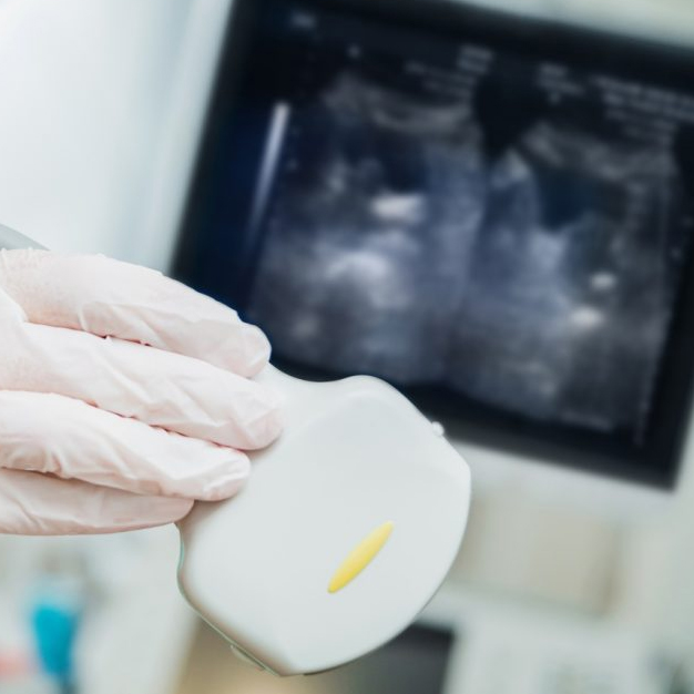
How it’s done: You will lie down on an examination table. A clear, water-based gel is applied to your skin over the area to be examined. A sonographer then gently presses and moves a handheld device (transducer) over your skin. The transducer sends out sound waves and receives their echoes, which are converted into images on a monitor.
Time: 20-60 minutes total.